
CT vs MRI: Key Differences Explained
Health ArticlesOct 14, 2026
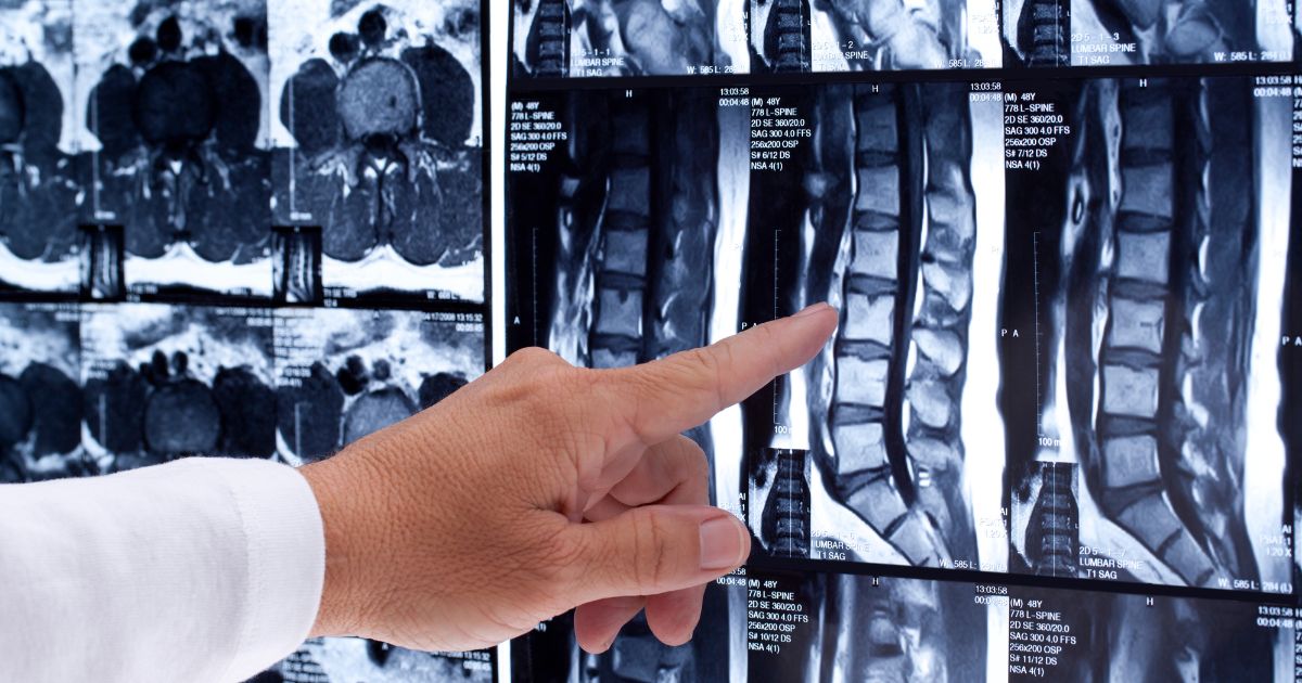
Medical Imaging: Understanding CT and MRI Scans
When your doctor orders an imaging test, it’s perfectly normal to have questions. Both CT scans and MRI scans provide a way for doctors to visualize the internal workings of the body without the need for invasive procedures. However, they are distinct modalities with differing methods of operation and suitability for various conditions. It is worth noting that a CT scan is also referred to as a CAT scan.
Summary
- CT scans employ X-rays, which means they involve exposure to radiation. They are quick and are often preferred for emergency situations, assessing trauma, and visualizing bone structures.
- MRI scans, on the other hand, utilize powerful magnets and radio waves, thereby eliminating radiation exposure. They are more adept at capturing details related to soft tissues, the brain, and spinal cord but generally require a longer duration to conduct and may pose challenges for patients with certain implants or in confined spaces.
- There isn't a universally superior option; the choice between CT and MRI depends on the specific condition that needs evaluation, and both modalities can involve the use of contrast agents to enhance visibility.
- Furthermore, safety considerations and logistics differ markedly between the two. While CT scans involve radiation exposure, MRI scans often have lengthier wait times for scheduling.
What is the Difference Between a CT Scan and an MRI?
Mechanisms of Operation
- CT (Computed Tomography): This technique employs X-rays captured from multiple angles to construct cross-sectional images of the body.
- MRI (Magnetic Resonance Imaging): This approach relies on the use of strong magnetic fields and radiofrequency waves to generate high-resolution images.
In essence, CT scans introduce radiation exposure, while MRI scans do not.
Experience During Scanning and Duration
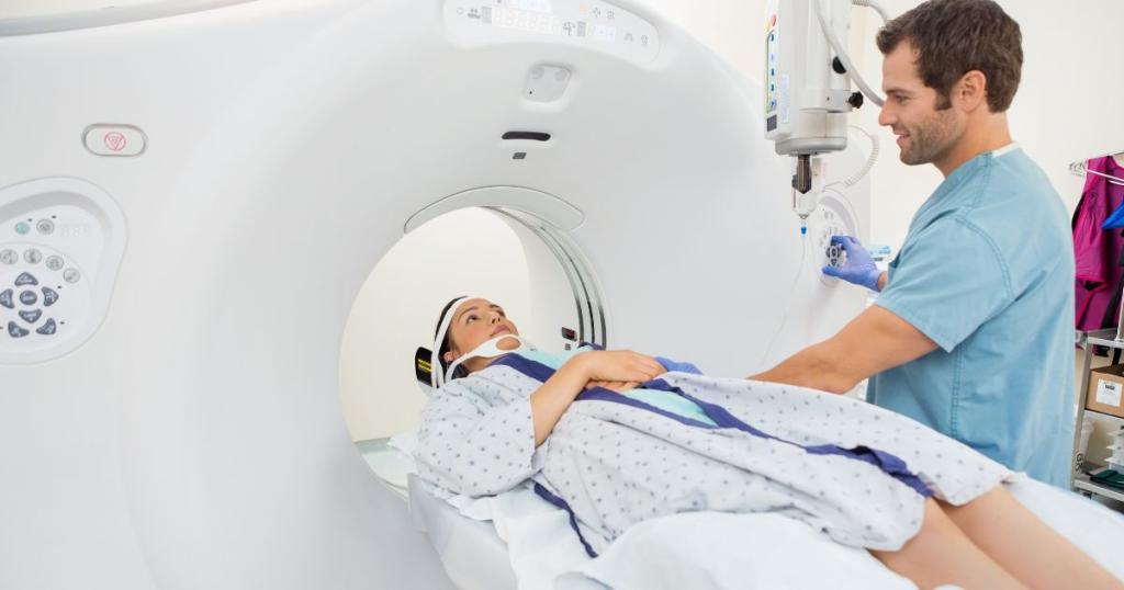
- CT scans are typically rapid, often completed within 5 to 10 minutes, which makes them a common choice in emergency medicine.
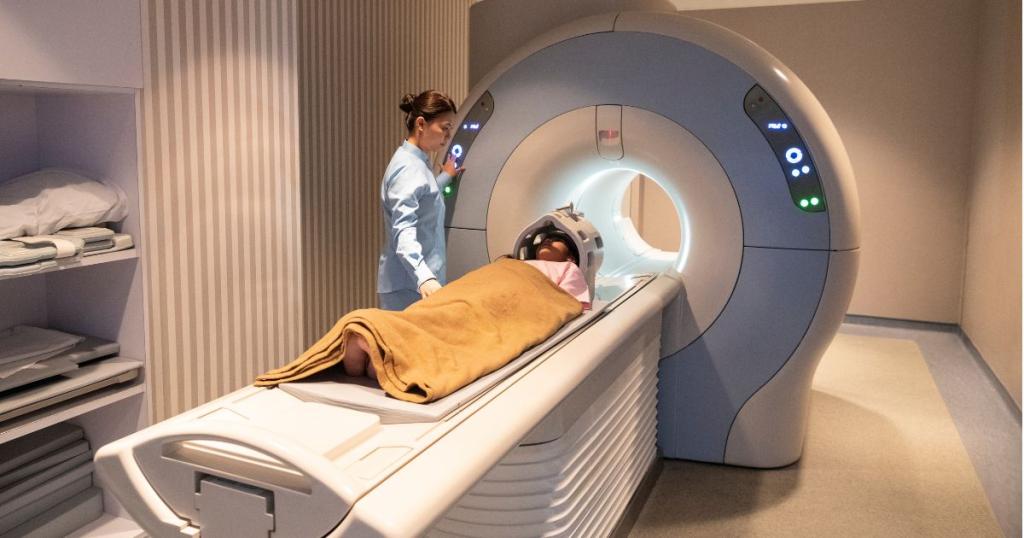
- In contrast, Orthopedic MRI scans may take approximately 30 minutes and require patients to lie within a longer, narrower tube.
- If you experience anxiety in tight spaces, it is important to communicate this to the imaging staff. Many facilities offer music, ear protection, or anxiolytic medication to help ease any discomfort during the procedure.
Determining the Better Option: CT or MRI?
There is no definitive answer as to which test is superior; the decision is based on the specific circumstances and clinical needs.
Scenarios Favoring CT Scans
- Emergencies and Trauma: The speed of CT scans makes them invaluable in urgent situations such as car accidents, severe falls, or sudden, acute symptoms.
- Assessment of Bleeding: CT scans are particularly effective in quickly identifying bleeding in critical scenarios.
- Bone Imaging: CT technology excels at visualizing fractures and providing detailed images of bone structures.
Situations Where MRI is Preferable
- Soft Tissue Assessment: MRI scans offer superior detail concerning muscles, ligaments, tendons, cartilage, spinal discs, and various nerve-related issues.
- Neurological Imaging: For evaluating conditions of the brain and spine that may be less apparent on CT, MRI is often the preferred tool.
CT vs MRI in Orthopedics
Orthopedic conditions often necessitate detailed imaging to diagnose injuries and assess structural integrity. Both CT scans and MRIs are valuable tools in the orthopedic field, providing complementary information depending on the specific circumstances.
CT Scans in Orthopedics
CT scans are particularly effective for evaluating bone structures due to their ability to produce detailed cross-sectional images. They are commonly used for:
- Fracture Assessment: CT scans can visualize complex fractures that may be difficult to interpret on standard X-rays, especially in areas like the pelvis, spine, and joints.
- Bone Tumors: They help in assessing bone tumors or lesions, determining their size, and investigating whether they have invaded surrounding tissues.
- Post-Surgical Evaluations: After orthopedic surgery, CT scans can be used to evaluate the positioning of hardware such as screws and plates or to check for complications such as infections or non-union of fractures.
MRI in Orthopedics
MRI is essential for assessing soft tissues in orthopedic patients. It provides high-resolution images of ligaments, tendons, cartilage, and muscles, making it suitable for:
- Tendon and Ligament Injuries: MRI is the preferred imaging modality for diagnosing tears in structures such as the anterior cruciate ligament (ACL) or rotator cuff in the shoulder.
- Cartilage Damage: It can clearly visualize cartilage wear and degeneration, especially in the knee joint, helping assess conditions like osteoarthritis.
- Bone Marrow Edema: MRI is exceptional at detecting bone marrow edema, which can indicate inflammatory conditions, stress fractures, or osteonecrosis.
Complementary Use
In many cases, orthopedic specialists may employ both modalities to obtain a comprehensive view of a patient's condition. For instance, a CT scan might be used to assess a complex fracture, while an MRI could subsequently evaluate soft tissue involvement around the injury. This dual approach aids in creating an effective treatment plan tailored to the patient's needs.
By understanding the specific advantages of CT scans and MRIs in orthopedics, healthcare providers can make informed decisions regarding imaging and treatment strategies for various musculoskeletal conditions.
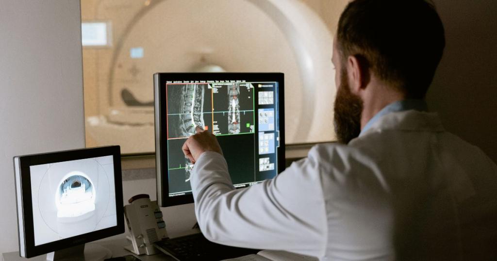
Safety Considerations and Preparation
Radiation Exposure
- CT scans involve ionizing radiation. Physicians carefully assess the benefits against the risks associated with this exposure and strive to minimize unnecessary scans.
- MRI scans, conversely, do not utilize radiation.
Considerations Regarding Metal and Implants
- Due to MRI’s reliance on strong magnets, certain implants, including specific types of pacemakers or metallic fragments, may render the procedure unsafe.
- Always inform the imaging personnel of any implants or prior surgical interventions.
Use of Contrast Agents
- Both CT and MRI scans sometimes necessitate the administration of contrast agents to improve image quality.
- Notify your healthcare team if you have a history of reactions to contrast media or if you suffer from renal impairment.
Cost, Comfort, and Scheduling Concerns
- In the Hampton Roads region, MRI and CT scans used for orthopedic imaging typically fall within a similar cost range.
- The MRI experience occurs in a tube-like structure that may be constraining. If claustrophobia is a concern, inquire about options for music, ear protection, or sedation.
Conclusion
The choice between CT and MRI is not straightforward; both have unique advantages depending on the clinical context. CT scans are typically expedited and frequently used for emergent evaluations, traumas, and bony anomalies. Conversely, MRI scans, while taking longer to perform, generally provide superior insights into soft tissue and complex neurological conditions. Ultimately, your physician will recommend the modality that aligns best with your symptoms and medical history.
Need Imaging Services? We're Here to Help.
Whether your physician recommends an MRI, CT scan, or another diagnostic imaging study, our experienced team is committed to providing convenient, high-quality care close to home. Learn more about the imaging services available at Tidewater Orthopaedics by visiting our Imaging Center page.
Ready to schedule an appointment? Click the link below to request an appointment with our team.
Q&A
What are the basic differences between CT scans and MRI?
Short answer: CT scans utilize X-rays to create slice images and involve ionizing radiation, typically completed within 5 to 10 minutes. MRI employs strong magnetic fields and radio waves without radiation, requiring approximately 30 minutes, conducted within a narrower tube structure.
Under what circumstances might my doctor opt for a CT scan over an MRI, and vice versa?
Short answer: CT scans are often preferred for acute medical situations and trauma due to their speed, excellent bone detail, and ability to detect internal bleeding. MRI is favored for evaluations requiring detailed visualization of soft tissues, including musculature, ligaments, and complex joint or spinal issues.
How are CT scans and MRIs utilized in orthopedics?
Short answer: In orthopedics, CT scans are utilized to assess bone integrity, visualize complex fractures, evaluate post-surgical outcomes, and detect bone tumors. On the other hand, MRIs are essential for imaging soft tissues, effectively diagnosing tendon and ligament injuries, cartilage damage, and conditions like bone marrow edema. Both modalities complement each other, allowing healthcare providers to develop comprehensive treatment plans based on the nature of the orthopedic condition.
Can CT scans confirm cancer, or are additional tests necessary?
Short answer: CT scans can identify masses and suspicious areas and are extensively utilized for tumor detection and assessment of cancer spread in the chest or abdomen. However, imaging results alone do not confirm cancer, necessitating follow-up imaging studies, additional scans, or biopsy procedures for a definitive diagnosis.
What key safety, comfort, and preparation issues should I be aware of regarding these imaging procedures?
Short answer: CT scans utilize ionizing radiation; thus, doctors carefully consider the risks associated with repeated exposure. MRI employs strong magnetic fields, which can pose risks for individuals with certain implants. Inform the imaging team of any prior surgeries or implants. Some scans require the use of contrast agents, so disclose any history of contrast reactions or kidney disease. MRI scans tend to take about 30 minutes and occur in a confined tube.
Sources
- RadiologyInfo.org. "Body MRI". https://www.radiologyinfo.org/en/info/bodymr
- RadiologyInfo.org. "Body CT". https://www.radiologyinfo.org/en/info/bodyct
- MedlinePlus (National Library of Medicine). "MRI Scans". https://medlineplus.gov/mriscans.html
- U.S. Food & Drug Administration (FDA). "Computed Tomography (CT)". https://www.fda.gov/radiation-emitting-products/medical-x-ray-imaging/computed-tomography-ct
